
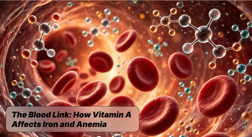
The Blood Link: How Vitamin A Affects Iron and Anemia. Persistent fatigue, brain fog, and low physical stamina often push people toward standard blood tests that flag low ferritin or sub-optimal hemoglobin levels. The universal reflex in modern wellness is to immediately supplement high doses of oral iron to combat this exhaustion. However, isolated mineral supplementation frequently hits a frustrating dead end, causing gastrointestinal distress without meaningfully shifting circulating red blood cell counts. The human body does not process vital nutrients in clean isolation, but rather through intricate biological feedback loops where cofactors dictate outcomes.
In the context of hematology, vitamin A affects iron and anemia by functioning as a biochemical key that unlocks systemic mineral utilization. Without adequate retinoids circulating in blood plasma, dietary minerals remain functionally inert, trapped inside storage tissues instead of driving vital daily cellular respiration and tissue oxygenation across every organ system.
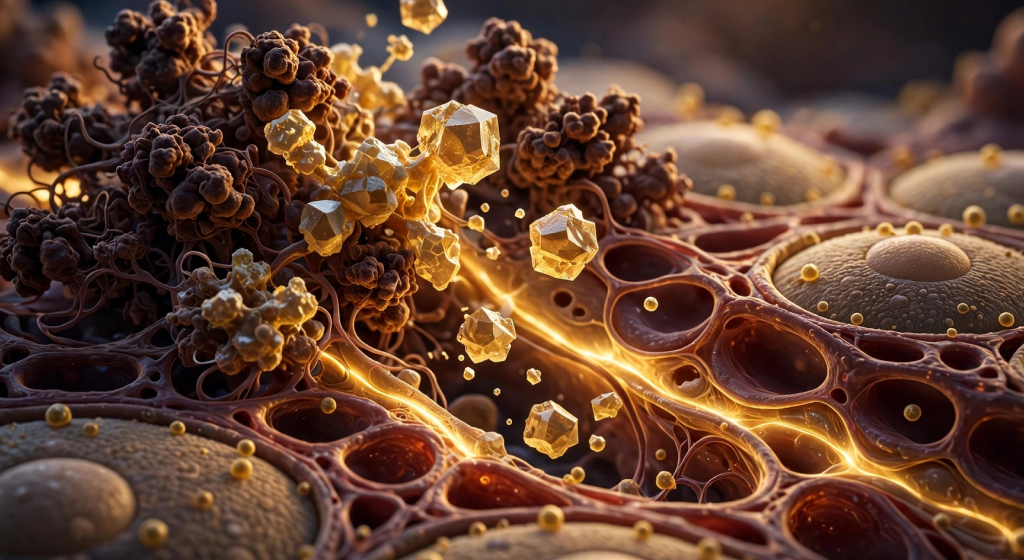
Understanding this biological interplay requires zooming out to look at whole-body nutrient status rather than chasing a single lagging marker. Many individuals diagnosed with refractory iron deficiency are surprised to discover that their tissues actually hold sufficient total iron that simply cannot reach functional pathways. Nutritional science demonstrates that a severe shortage of bioavailable retinol directly mirrors the cellular symptoms of systemic mineral starvation. When the body lacks sufficient retinoid reserves, the production of vital transport proteins stalls, enzymatic activation falters, and healthy red cell maturation slows to a crawl. Biohackers and wellness enthusiasts seeking peak daily energy must therefore evaluate the entire micronutrient matrix rather than treating blood panels with isolated, single-molecule interventions:
- Retinoids govern gene transcription for the transport machinery that makes daily cellular mineral assimilation possible.
- Trapped mineral stores cannot mobilize into circulation when critical fat-soluble cofactors fall below physiological thresholds.
- Targeted biohacking strategies prioritize dual-nutrient synergy over aggressive single-compound megadosing protocols.

Decoding Anemia Beyond Simple Iron Scarcity
Classic clinical anemia is technically defined as a reduction in circulating red blood cells or a drop in total hemoglobin concentration below normal reference ranges. While iron deficiency anemia represents the most widely recognized variant globally, clinical research confirms that micronutrient imbalances rarely occur in neat, singular silos. Insufficient dietary intake of retinol creates a distinct functional state known as vitamin A deficiency anemia, which frequently gets misdiagnosed as pure mineral exhaustion due to overlapping clinical presentations. When functional retinoids run low, bone marrow stem cells lose the essential biochemical signals required to differentiate into healthy, oxygen-carrying erythrocytes. The resulting physiological bottleneck produces identical symptoms: cold extremities, rapid heart rates during light exertion, persistent midday lethargy, and compromised cognitive clarity.
Addressing persistent fatigue demands that you look closely at cellular architecture and biological signaling pathways. Standard medical checks focus primarily on serum ferritin, which measures static mineral storage but tells you very little about active mobilization dynamics. If retinol is missing from the cellular environment, systemic recycling grinds to a halt, leaving the body functionally anemic despite normal tissue reserves. This hidden deficiency pattern explains why conventional mineral pills frequently fail to produce lasting vitality improvements, leaving individuals frustrated and symptomatic despite months of diligent daily intake. Broadening your diagnostic awareness reveals that optimizing oxygen transport across the vascular tree requires addressing the signaling lipids that govern red cell creation from the start:
- Functional anemia often reflects poor cellular signaling rather than an absolute lack of ingested dietary minerals.
- Erythrocyte differentiation in the bone marrow relies heavily on active retinoic acid receptor activation.
- Misinterpreting storage biomarkers leads to ineffective supplementation routines that overlook critical fat-soluble coenzymes.
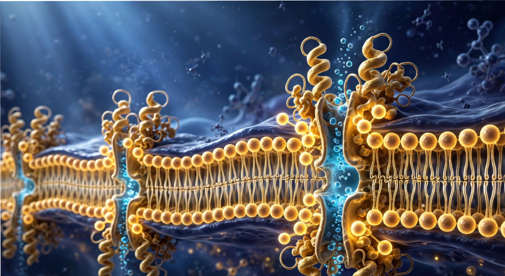
The Mobilization Mechanism: Freeing Trapped Iron Stores
The liver serves as the primary metabolic warehouse for systemic mineral reserves, packaging them inside a specialized spherical protein complex known as ferritin. Under normal conditions, these reserves are systematically mobilized into systemic circulation whenever peripheral tissues require fresh building blocks for hemoglobin synthesis. However, this liberation process is strictly regulated by retinoid-dependent signaling pathways that govern hepatic transport channels. Research into micronutrient transport reveals that vitamin A mobilizes stored iron from liver hepatocytes directly into the bloodstream, making it readily accessible for functional tissue needs. Without sufficient circulating retinol, hepatic mineral release becomes sluggish, effectively locking vast stockpiles of vital raw materials inside organ tissues where circulating blood cells cannot reach them.
This biological trapping mechanism produces a confusing clinical picture where a person exhibits low serum iron alongside paradoxically elevated or normal ferritin levels. When retinoic acid binds to specific nuclear receptors within liver cells, it triggers the enzymatic machinery responsible for disassembling ferritin storage cages and handing minerals off to circulating transport proteins. Without this vital biochemical handshake, stored reserves stagnate, fostering cellular stress within the liver while peripheral tissues starve for oxygen. Restoring adequate retinoid levels acts like opening a biological dam, rapidly releasing dormant mineral assets back into circulation without requiring high-dose mineral supplements that could irritate your gut:
- Retinol triggers the transcriptional release of hepatic reserves into active vascular circulation when cellular demand spikes.
- Static ferritin deposits remain biologically inaccessible to bone marrow tissues without active retinoid-driven signaling cascades.
- Optimizing fat-soluble vitamins restores natural mineral flux, resolving functional deficiencies safely without causing digestive upset.
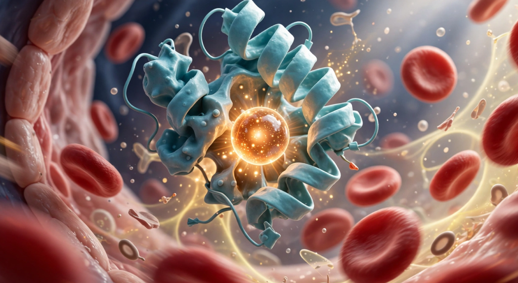
Regulating Hepcidin and Systemic Mineral Flow
Systemic iron homeostasis is meticulously governed by hepcidin, a small peptide hormone synthesized by the liver that acts as the master circuit breaker for mineral metabolism. When hepcidin levels rise in response to systemic inflammation or infection, it binds to and degrades ferroportin, the sole cellular exporter responsible for moving minerals from intestinal enterocytes and macrophages into blood plasma. High hepcidin essentially locks cellular gates, shutting down intestinal absorption and stranding recycled minerals inside immune cells. In this delicate hormonal environment, retinol modulates hepcidin expression, dampening excessive production of this inhibitory hormone and preserving smooth mineral transit through critical vascular checkpoints so oxygen delivery remains optimal.
By keeping systemic hepcidin levels balanced and low, bioavailable retinol prevents the functional blockades that characterize the anemia of chronic inflammation. Sub-clinical systemic stress, intense physical training, and gut dysbiosis can all trigger localized immune cascades that drive hepcidin upwards, rapidly cutting off tissues from their vital oxygen-carrying nutrients. Retinoids intervene directly at the genomic level, interacting with cellular signaling cascades to suppress unnecessary hepcidin transcription and keep cellular ferroportin exporters open. This hormonal regulation ensures that daily dietary nutrients enter circulation seamlessly rather than being excreted or sequestered inside damaged gut linings:
- Hepcidin acts as a biological master switch, shutting down systemic mineral absorption whenever systemic inflammation spikes.
- Active retinoic acid downregulates hepcidin gene expression, keeping cellular export doorways fully open and functional.
- Maintaining low, stable hepcidin levels protects active fitness enthusiasts from exercise-induced functional mineral lockups.
Transferrin and Cellular Mineral Delivery
Once dietary or recycled minerals successfully exit cellular storage sites, they cannot travel through the vascular system in an unbound, free-floating state. Free circulating minerals induce violent oxidative stress, generating destructive reactive oxygen species through classic Fenton chemistry that tears through delicate endothelial vessel walls. To prevent this severe vascular damage, the body relies on transferrin, a high-affinity glycoprotein that securely captures circulating minerals and shuttles them safely to distant target organs. Here again, the vital nutrient connection shows its power: vitamin A stimulates transferrin synthesis within liver hepatocytes, ensuring that the vascular system maintains ample carrier capacity to shepherd minerals safely across blood vessels to ravenous target tissues.
When hepatic retinol stores run low, circulating transferrin levels drop noticeably, severely impairing the body’s total iron-binding capacity and slowing systemic transit times. Even if minerals successfully escape cellular storage depots, a shortage of transferrin creates an infrastructural bottleneck where nutrients cannot reach the erythroblasts in bone marrow that build red blood cells. By supporting robust transferrin transcription and membrane receptor density, optimal retinol levels ensure that every mobilized molecule reaches its metabolic destination quickly and without inducing damaging oxidative cascades along arterial pathways:
- Transferrin prevents circulating mineral toxicity by shielding vascular walls from catastrophic free-radical oxidative damage.
- Liver synthesis of transferrin transport carriers depends directly on healthy, uncompromised fat-soluble retinoid reserves.
- Elevated carrier capacity accelerates the targeted delivery of raw building materials to developing red marrow cells.

Bone Marrow Erythropoiesis and Oxygen Delivery
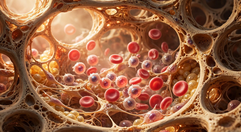
The manufacturing of new red blood cells—a tightly choreographed physiological process known as erythropoiesis—occurs continuously deep within the spongy interior of human bone marrow. Hematopoietic stem cells must undergo a precise, multi-stage developmental journey, transforming from uncommitted progenitor cells into fully mature, oxygen-rich red blood cells loaded with functional hemoglobin. Scientific investigations confirm that erythropoiesis requires active vitamin A to guide this rapid cellular differentiation and prevent premature cell death during early developmental stages. Retinoic acid binds to specialized nuclear receptors inside the bone marrow microenvironment, directly upregulating genes that coordinate hemoglobin synthesis and protect young red blood cells from oxidative stress.
When bone marrow tissues are deprived of adequate retinoids, the assembly line of blood cell production experiences high rates of cellular failure and developmental arrest. Immature blood cells often die prematurely inside the marrow matrix, leading to a marked decrease in functional reticulocyte counts entering peripheral circulation each day. Furthermore, retinoid signaling works in tight physiological harmony with erythropoietin, the primary kidney hormone that stimulates red cell creation during periods of low oxygen. Maintaining optimal levels of this master fat-soluble vitamin guarantees that your bone marrow receives both the genetic instructions and the structural cofactors required to produce robust, flexible red blood cells capable of circulating through narrow capillary networks:
- Retinoic acid nuclear receptors directly activate the genetic blueprints for functional hemoglobin protein synthesis.
- Bone marrow progenitor cells require sustained retinoid signaling to mature safely without undergoing programmed cell death.
- Synergy between kidney-derived erythropoietin and dietary retinoids maximizes functional oxygen-carrying capacity during athletic exertion.

Final Thoughts: “The Blood Link: How Vitamin A Affects Iron and Anemia”
True biohacking is never about chasing isolated biomarkers with aggressive, single-compound megadoses; it is about respecting the intricate regulatory networks that govern human physiology. When fatigue, sluggish recovery, or stubborn lab panels point to low oxygen-carrying capacity, looking at iron alone offers only half the equation. The biochemical bridge between fat-soluble retinoids and mineral homeostasis proves that the body requires the right genetic signals, transport carriers, and hormonal balance to put vital nutrients to work. By ensuring adequate, bioavailable retinol intake alongside strategic lifestyle habits that keep systemic inflammation low, you dismantle the hidden bottlenecks that keep essential minerals locked away in storage.
Moving forward, shift your focus from merely accumulating raw materials to optimizing their functional utilization across every organ system. Elevating your energy architecture begins with comprehensive diagnostics that evaluate serum retinol, transport capacity, and inflammatory markers as an interconnected team. Prioritize whole-food micronutrient synergy, pair non-heme meals with fat-soluble cofactors, and treat nutrient absorption as a dynamic cellular conversation rather than a static transaction. When your biochemical machinery has both the fuel it needs and the signaling cofactors to direct it, mitochondrial respiration flourishes, tissue oxygenation peaks, and resilient daily vitality becomes your natural baseline:
- Sustainable vitality relies on cellular nutrient mobilization rather than isolated, high-dose mineral saturation.
- Prioritizing fat-soluble retinoid reserves ensures your body possesses the signaling tools needed to govern systemic mineral flow.
- Optimizing the internal micronutrient matrix unlocks peak oxygen delivery, physical endurance, and sustained mental clarity.