
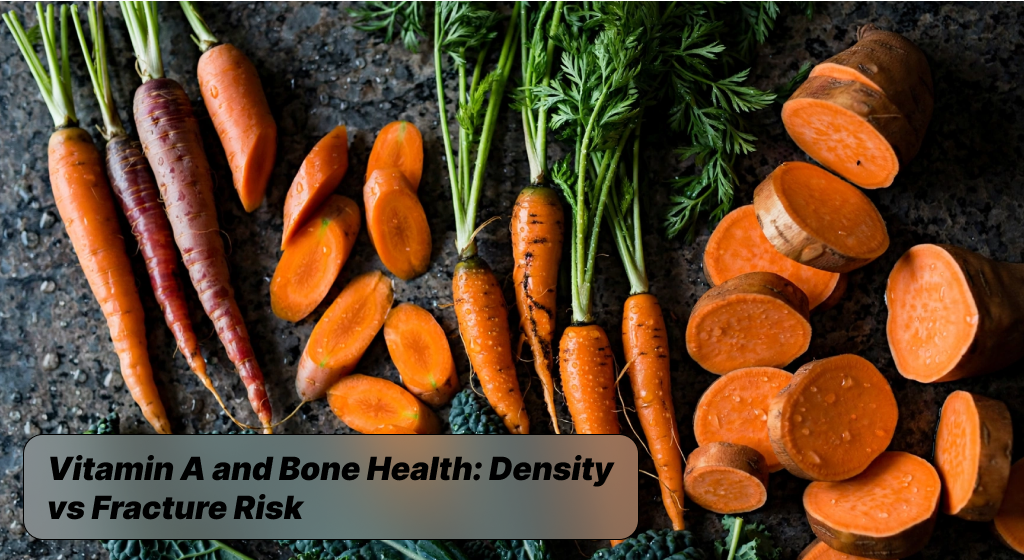
Vitamin A and Bone Health: Density vs Fracture Risk. Vitamin A plays a fundamental role in maintaining skeletal integrity, yet its biological relationship with human bones operates on a strict concentration curve. As a fat-soluble retinoid, it actively directs gene transcription inside marrow stromal cells, telling immature precursor cells whether to form structural matrix tissue or specialize into other cell lineages. When systemic levels stay within an optimal physiological range, circulating retinol acts as an indispensable coordinator of standard tissue repair.
However, human skeletal architecture does not respond to retinoids in a linear fashion where higher intakes guarantee stronger scaffolding. Instead, the skeletal system treats both insufficient supply and chronic excess as significant disruptors of natural collagen deposition and mineralization. Maintaining stable circulation ensures baseline connective integrity without triggering stress responses inside the mineralized osteoid tissue.
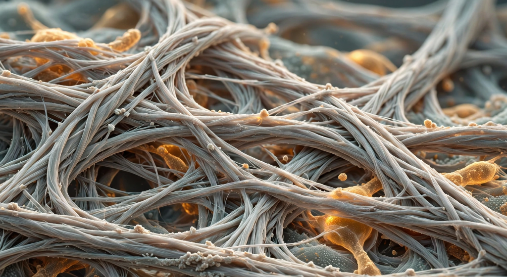
The core paradox emerges when comparing laboratory bone scans against actual clinical outcomes in everyday movement. A dense osteoid framework does not automatically translate to a resilient skeleton if structural microdamage accumulates unchecked over time. Retinoids significantly influence cellular signaling across the periosteum and endosteum, altering the internal balance required to handle sudden mechanical shear stresses.
Individuals focused on cellular longevity and targeted micronutrient optimization must recognize that bone quality involves far more than simple mineral mass measurements. Achieving robust skeletal protection demands a clear distinction between internal mineral accumulation and the functional mechanics of connective tissue elasticity. Retinoids drive this entire matrix environment through continuous epigenetic signaling across your skeletal architecture.
- Bone mineral mass reflects calcium deposit volume but completely overlooks structural brittleness caused by accelerated cellular turnover.
- Cellular differentiation depends on continuous active retinoid exposure to guide osteoblast lineages toward mature collagen production.
- Systemic equilibrium safeguards internal trabecular struts against premature resorptive remodeling triggered by fluctuating serum retinol levels.

Cellular Dynamics Between Osteoclasts and Osteoblasts
Skeletal tissue continuously regenerates itself through coordinated remodeling led by matrix-synthesizing osteoblasts and specialized matrix-clearing osteoclasts. Specialized receptors inside osteogenic precursor cells absorb circulating retinol and convert it locally into all-trans retinoic acid, directly activating nuclear transcription pathways. This regulatory cascade stimulates initial osteoblast activity, producing the flexible Type I collagen fibers that form the structural foundation of the skeletal frame.
Without adequate signaling from active retinoids, osteoblasts fail to assemble organized organic scaffolds, resulting in disorganized osteoid tissue incapable of binding essential minerals properly. Consistent retinoid availability supports stable basic cellular metabolism and maintains ongoing collagen synthesis during daily physical activities.
Problems emerge when high concentrations of retinoid molecules overstimulate basic osteoblast signaling cascades, prompting defensive paracrine reactions across the local microenvironment. Stressed osteoblasts quickly release receptor activator of nuclear factor kappa-B ligand, an essential chemical trigger known as RANKL. Elevated levels of RANKL bind directly to local pre-osteoclasts, forcing them to mature into aggressive, resorptive osteoclasts that clear calcified tissue far faster than standard rates.
This accelerated breakdown strips away cortical thickness along the outer skeletal shell while osteoblasts struggle to lay down fresh mineralized collagen fast enough. Once resorptive osteoclasts outpace matrix-depositing osteoblasts, the internal lattice of trabecular struts thins out, turning standard bone remodeling into an active vulnerability.
- Targeted gene expression prompts osteoblasts to lay down fresh structural protein scaffolds across the organic bone matrix.
- Signaling through RANKL activates specialized osteoclasts when elevated local retinoid concentrations exceed baseline skeletal needs.
- Disrupted cell balance speeds up natural tissue resorption, hollowing out internal struts faster than replacement osteoid can mineralize.

Bone Mineral Density Versus Trabecular Microarchitecture
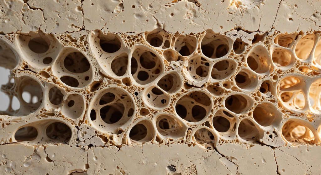
Standard clinical diagnostics routinely measure bone mineral density via dual-energy X-ray absorptiometry scans, creating a two-dimensional snapshot of overall mineral mass. While DXA scans provide useful baseline metrics, they fail to reveal the precise spatial layout of internal trabecular microarchitecture. Bone tissue resembles a high-performance architectural bridge where lightweight crisscrossing struts distribute multidirectional mechanical stresses smoothly across the entire skeletal frame. A bone can hold abundant calcium mineral deposits and produce reassuring readings on two-dimensional density scans while concealing microstructural thinning within its load-bearing interior. Focusing exclusively on density numbers creates a false sense of skeletal security because it hides qualitative matrix defects.
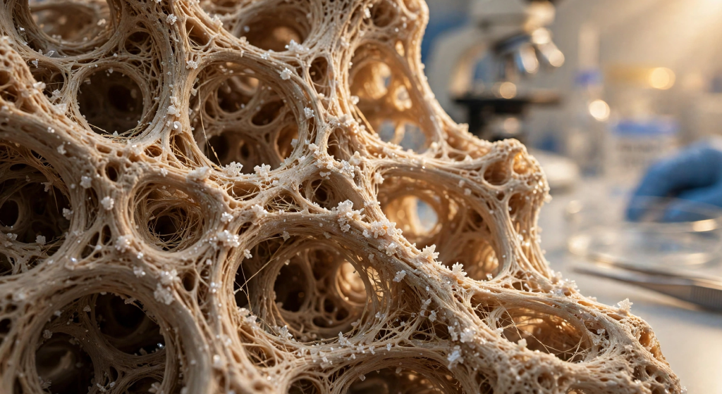
Excessive active retinoid consumption alters structural remodeling, preferentially attacking internal trabecular connections while leaving coarse mineral mass relatively intact. This selective cellular remodeling strips away delicate horizontal cross-struts, reducing complex mechanical connectivity throughout internal spongy tissue compartments. As these structural bridges vanish, the remaining vertical pillars carry significantly higher localized strain whenever the skeleton experiences sudden impacts or torsional force.
The resulting bone structure exhibits high structural rigidity alongside severe microstructural brittleness, making the framework susceptible to failure without any prior warning drops on standard density charts. Real skeletal durability demands intact geometric distribution of internal matrix fibers, not just dense concentrations of static calcium salts.
- Standard density scans measure bulk mineral distribution across flat planes while missing microscopic fractures hidden inside spongy bone.
- Trabecular connections distribute sudden directional impact forces smoothly across internal skeletal bridges to prevent sudden structural failure.
- Microstructural brittleness develops when horizontal struts disappear, leaving isolated vertical columns vulnerable to shear stresses.
Decoupling Laboratory Density from Real Fracture Risk
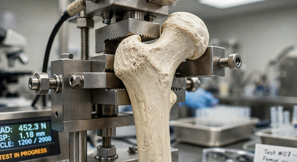
The critical medical distinction between measurable bone mineral density and actual fracture risk becomes stark when assessing long-term retinoid exposure across patient populations. Clinical cohorts with consistently high serum retinol levels frequently experience hip and femoral neck fractures despite presenting completely average T-scores during diagnostic imaging. This statistical contradiction shows that fracture events stem primarily from material fatigue and compromised structural geometry rather than simple mineral shortages. When bones lose structural compliance, they fail to absorb kinetic shock waves produced during standard daily activities like walking down stairs or navigating uneven terrain. The resulting kinetic shock waves propagate directly into brittle mineralized planes, initiating microscopic shear fractures.
Investigating real fracture risk requires evaluating the mechanical elasticity of the underlying collagen matrix alongside traditional mineral accumulation metrics. Retinoid toxicity subtly degrades collagen cross-linking profiles, creating a stiffened, glassy framework that shatters under sudden torsional strain instead of flexing harmlessly. Furthermore, cortical thinning around critical femoral neck zones concentrates baseline body weight across an increasingly narrow outer shell. Because conventional laboratory evaluations focus entirely on gross mineral quantities, they fail to detect this dangerous loss of functional bone quality. Protecting skeletal health requires prioritizing qualitative bone resilience and material elasticity over arbitrary targets for static mineral weight.
- Elevated fracture rates regularly appear in long-term observational trials despite completely normal baseline diagnostic density scores.
- Impaired material compliance stops collagen networks from bending naturally under sudden rotational stresses and everyday mechanical impacts.
- Cortical bone thinning concentrates mechanical weight onto narrow skeletal margins, significantly increasing the probability of sudden hip fractures.
Navigating the Competitive Receptor Crosstalk with Vitamin D
The biological relationship connecting Vitamin A and Vitamin D3 functions through an intricate, competitive balance inside cellular nuclei throughout the skeletal frame. Both micronutrients utilize nuclear receptor proteins that must form a functional physical heterodimer with the Retinoid X Receptor, or RXR, to begin transcription. When calcitriol activates its specialized Vitamin D Receptor, it seeks out an unattached RXR molecule to access specific DNA response elements that control structural calcium absorption.
If cellular fluid contains excessive quantities of active retinoic acid, monomeric RAR units quickly monopolize available RXR cofactors, blocking calcitriol complexes from binding properly. This direct competitive crosstalk effectively silences Vitamin D transcriptional pathways, even when patients display healthy circulating calcidiol concentrations on standard laboratory blood panels.
This nuclear receptor blockage creates a state of localized tissue resistance where intestinal cells fail to synthesize calcium-binding transport proteins efficiently. Consequently, serum calcium levels decline slightly, triggering a compensatory release of parathyroid hormone to scavenge required minerals directly from healthy bone stores. At the same time, maintaining a balanced ratio between retinoic acid and active Vitamin D generates synergistic protective benefits across marrow stromal tissues.
Balanced receptor dimerization promotes healthy alkaline phosphatase synthesis, ensuring that newly deposited collagen beds mineralize evenly without precipitating harmful amorphous crystals. Supporting smooth skeletal remodeling requires maintaining balanced co-factor availability across cell nuclei rather than flooding cellular receptors with disproportionate doses of single isolated compounds.
- Competitive RXR binding prevents activated Vitamin D complexes from anchoring to target DNA strands when retinoic acid saturates receptors.
- Functional tissue resistance reduces intestinal calcium uptake mechanisms despite adequate circulating levels of basic Vitamin D substrates.
- Balanced receptor dimerization activates proper alkaline phosphatase expression, supporting clean mineral crystal distribution throughout mature matrix tissue.
The Protective Synergy of Menaquinone-7 and Balanced Mineralization
Integrating adequate Vitamin K2 provides an essential metabolic safeguard that prevents excessive retinoid signaling from degrading healthy skeletal framework tissue. While Vitamin A and Vitamin D collaborate to generate structural osteocalcin within active osteoblasts, this essential matrix protein remains biologically dormant upon secretion. Inactive osteocalcin floats uselessly through the extracellular fluid unless specific enzymes complete a critical carboxylation reaction requiring continuous menaquinone availability.
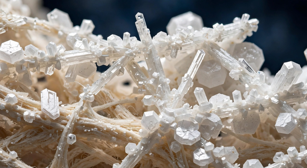
Once fully carboxylated by K2-dependent enzymes, osteocalcin develops a powerful electrostatic affinity for circulating calcium ions, pulling them into the collagen matrix. This precise biochemical handoff ensures that mobilized minerals integrate directly into crystalline hydroxyapatite plates rather than wandering freely into delicate vascular walls.
Without sufficient Vitamin K2, elevated retinoid levels simply accelerate the production of uncarboxylated, dysfunctional osteocalcin that cannot bind free calcium effectively. This metabolic failure leaves the newly formed organic matrix partially unmineralized while increasing free calcium concentrations inside systemic circulation. When combined with retinoid-driven osteoclast activity, this structural imbalance strips minerals from central load-bearing zones while accelerating soft-tissue calcification elsewhere in the body. Ensuring consistent dietary availability of long-chain menaquinones like MK-7 transforms basic retinoid signaling into stable, durable skeletal reinforcement. True skeletal optimization relies entirely on this coupled nutrient cascade to guide circulating minerals into their intended structural bone positions.
- Protein carboxylation transforms dormant matrix osteocalcin into a functionally active compound capable of binding ionic calcium securely.
- Controlled mineral placement directs circulating calcium crystals straight into structural bone matrix lattices instead of vulnerable arterial walls.
- Nutrient synergy safeguards eliminate uncarboxylated osteocalcin accumulation, protecting active bone remodeling zones from developing structural flaws.
Final Thoughts on “Vitamin A and Bone Health: Density vs Fracture Risk”
Navigating the delicate line between bone mineral density and real-world fracture resistance reveals a vital truth in nutritional biohacking: more is not inherently better. Skeletal health is an intricate, dynamic ecosystem governed by structural elasticity, geometric microarchitecture, and harmonious cellular signaling rather than sheer mineral volume. While preformed retinol remains an indispensable driver of cellular differentiation and immune function, unregulated systemic accumulation subtly shifts the remodeling balance toward trabecular degradation and microstructural brittleness. True longevity does not rely on megadosing single isolated compounds, but on mastering the biological dance of fat-soluble co-factors—ensuring that Vitamin D3, Vitamin K2, and bioavailable carotenoids work synergistically to guide minerals into their rightful crystalline matrices.
By shifting your nutritional strategy toward tightly regulated dietary precursors like beta-carotene, auditing supplement labels for hidden retinyl esters, and applying progressive axial loads through consistent strength training, you build a skeletal framework that is both mineral-dense and mechanically resilient. Protecting your framework against structural failure requires looking past static DXA scores and actively nurturing the living, flexible collagen networks beneath the surface. Real bone durability comes down to qualitative matrix strength, controlled receptor crosstalk, and sustained metabolic harmony, safeguarding your mobility and physical vitality across every decade of life.
- Prioritize matrix compliance alongside raw bone density to safeguard your skeletal system against unexpected shear fractures and torsional stress.
- Maintain co-factor balance by pairing moderate retinoid exposure with adequate Vitamin D3, MK-7, and essential mineral co-enzymes.
- Stimulate structural remodeling with purposeful mechanical loading to reinforce internal trabecular bridges for enduring, life-long mobility.