
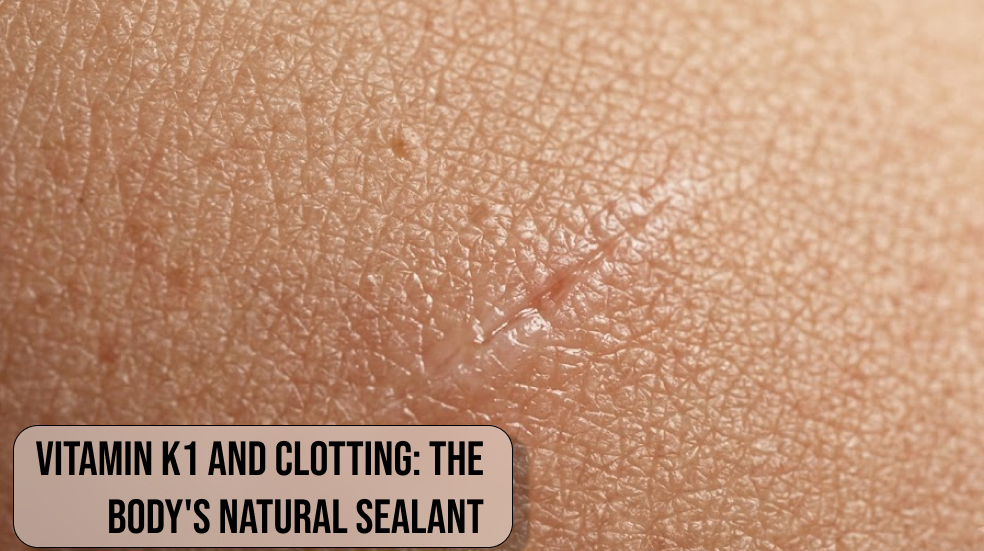
Vitamin K1 and Clotting: The Body’s Natural Sealant. When a microscopic cut or unexpected vascular injury occurs, the human body initiates an immediate, highly synchronized biochemical defense to stop liquid loss. At the very center of this essential protective reaction sits Vitamin K, a fat-soluble nutrient that functions as an emergency molecular sealant. While many vitamins support general cellular health over long timelines, Vitamin K1—scientifically designated as phylloquinone—acts as an instant catalyst for physiological repairs.
Without sufficient circulating amounts of Vitamin K, the vascular system loses its innate capacity to plug structural leaks, turning simple scratches into prolonged bleeding events. Biohackers and health enthusiasts often explore complex performance protocols, yet maintaining a fully functional hemostasis process through adequate Vitamin K intake remains the absolute baseline for structural resilience.
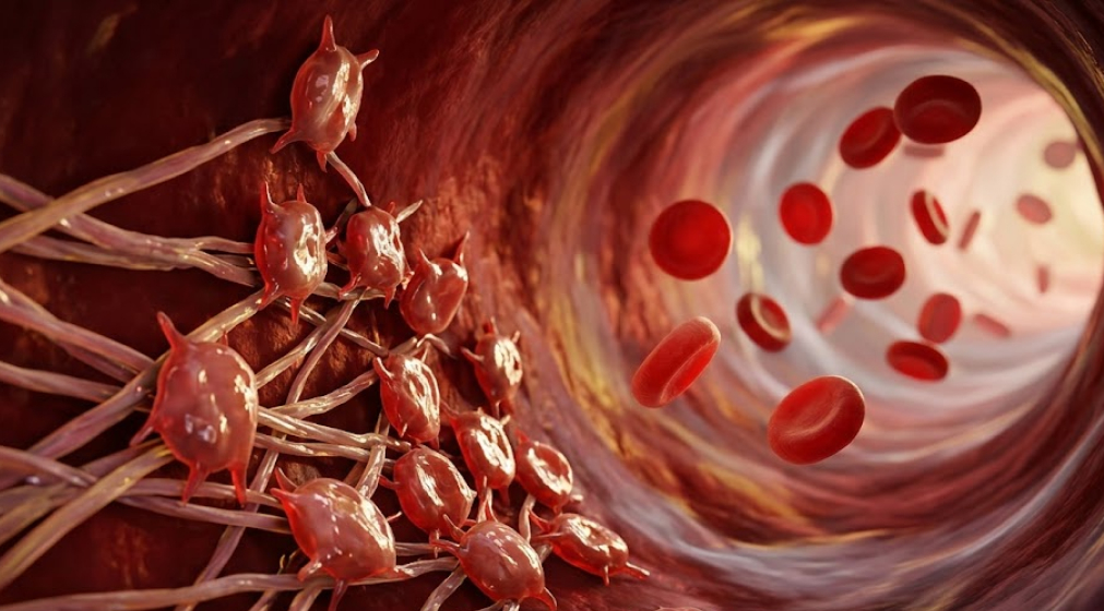
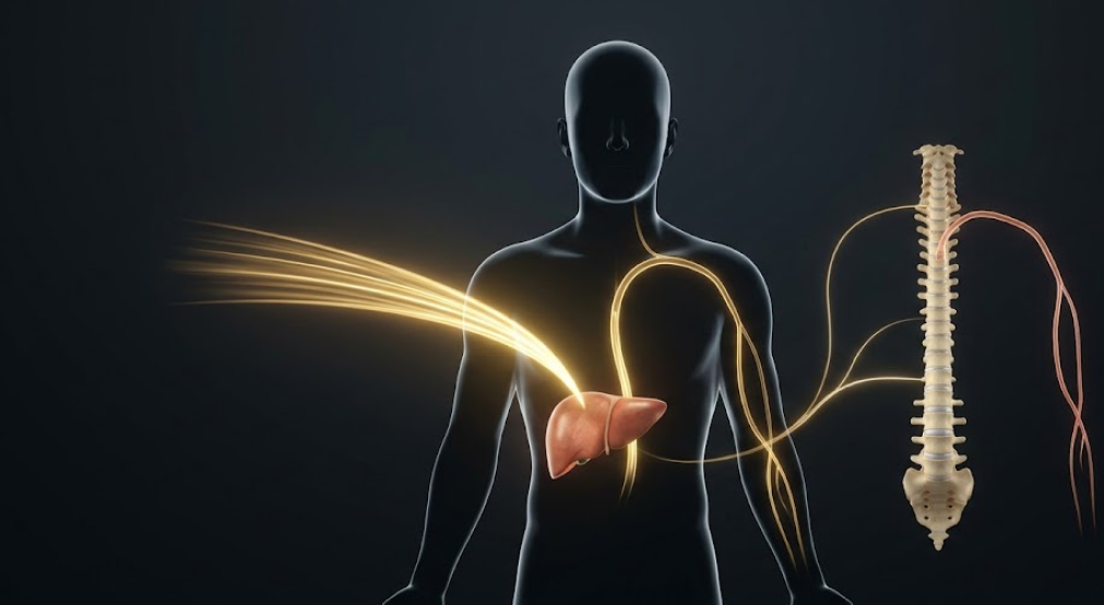
The mechanics of natural vascular sealing depend entirely on precise molecular timing and adequate liver reserves. Once phylloquinone absorption takes place inside the small intestine, this vital form of Vitamin K travels through lipid transport pathways straight into circulation. The liver rapidly intercepts these incoming nutrients to synthesize inactive precursor proteins that circulate quietly throughout the bloodstream. These inactive molecules wait for chemical alarm signals generated by damaged endothelial cells. When injury strikes, Vitamin K1 enables these dormant factors to transform into active enzymes almost instantly. This rapid biochemical conversion ensures that microscopic tears in capillaries and major blood vessels close securely, preserving vital fluid volume and maintaining overall cardiovascular balance.
- Rapid wound response: Triggers the immediate biochemical cascade required to stop bleeding at damaged vessel sites using essential Vitamin K factors.
- Hepatic nutrient accumulation: Moves directly to liver tissue after digestion to activate crucial proteins within the human coagulation system.
- Plant-derived dietary availability: Abundantly synthesized in dark green leafy vegetables, making daily fulfillment of phylloquinone simple and natural.
The Step-by-Step Biological Cascade of Hemostasis
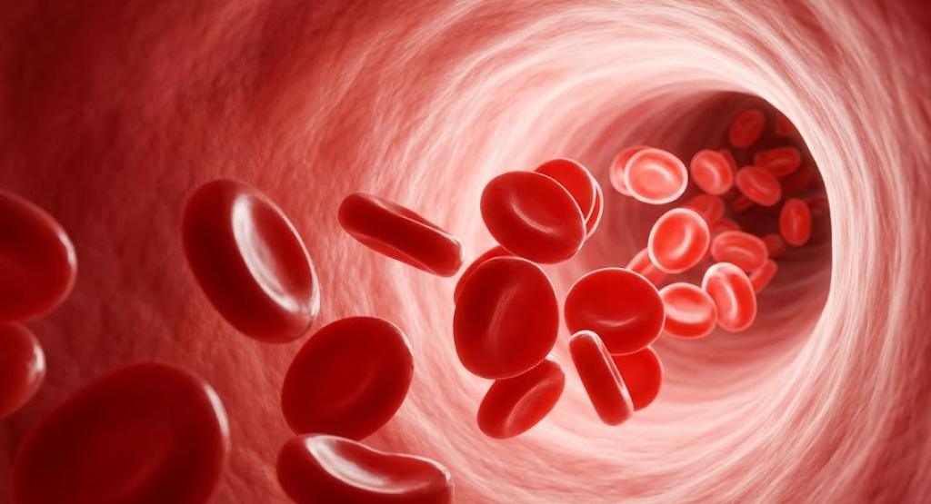
Halting internal or external bleeding is not a simple single-step chemical reaction, but rather a sophisticated biochemical domino effect known as the coagulation cascade. This multi-stage physiological mechanism ensures that blood remains smoothly fluid under normal conditions while quickly solidifying at localized sites of vascular injury. The entire protective sequence begins with immediate vascular constriction and platelet aggregation, where circulating cell fragments adhere to exposed collagen fibers at the injury site. However, a loose plug made entirely of platelets remains structurally vulnerable to normal blood pressure. To reinforce this temporary barrier, the body relies on clotting factor synthesis driven by Vitamin K, where sequential enzymatic reactions build a durable physical network.
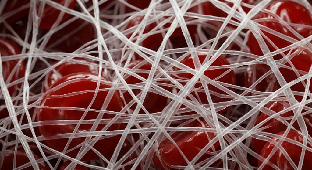
The ultimate objective of this delicate biological sequence is constructing a resilient fibrin mesh that securely captures red blood cells and seals the recovering tissue. Throughout this intricate process, various blood proteins transition from inactive states into active enzymes through specialized chemical modifications. Every single stage magnifies the next step, converting a tiny initial injury signal into thousands of active repair enzymes. Proper tissue repair requires strict spatial control so that the resulting clot remains strictly localized to damaged areas without blocking adjacent healthy vessels. Vitamin K1 acts as the crucial master regulator that allows these specialized proteins to attach properly to cell membranes, ensuring that blood clotting proceeds with complete spatial accuracy.
- Vascular constriction: Narrows damaged blood vessels immediately to reduce local blood flow and control physical pressure.
- Platelet plug formation: Assembles a sticky temporary physical barrier across damaged endothelial walls to prevent fluid loss.
- Fibrin network activation: Converts soluble fibrinogen into insoluble threads using Vitamin K dependent enzymes to complete structural sealing.
Molecular Action: Gamma-Glutamyl Carboxylation Explained
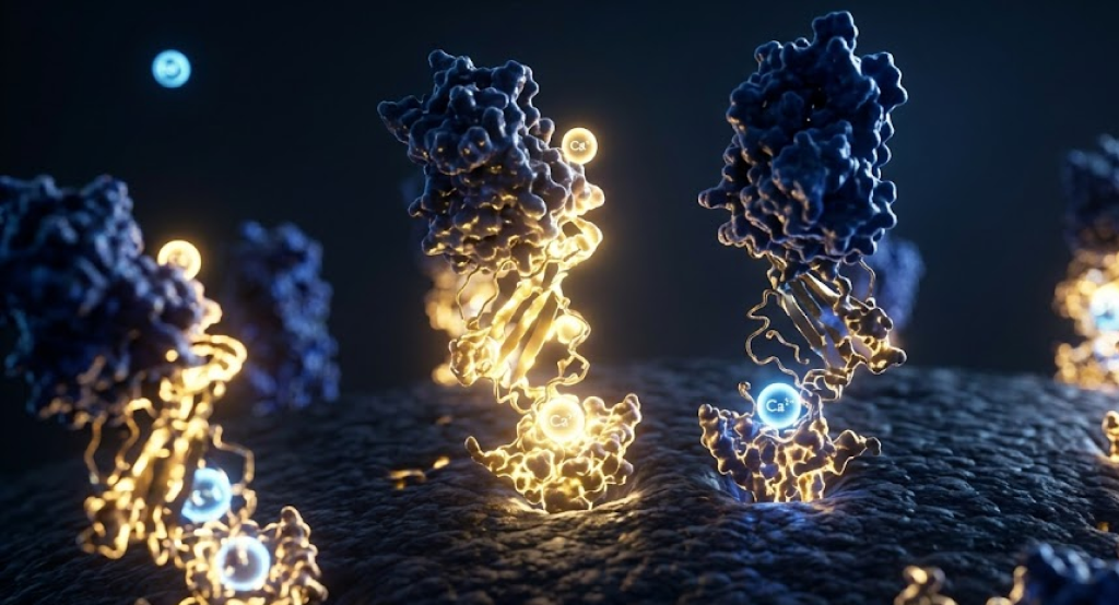
To truly appreciate how fat-soluble Vitamin K enables rapid seal formation, one must analyze a specific microscopic reaction called gamma-glutamyl carboxylation. Inactive clotting proteins manufactured by the liver contain specific glutamic acid residues that initially lack the negative electrical charge necessary to bind to blood vessel walls. Inside liver cells, a specialized enzyme known as gamma-glutamyl carboxylase utilizes Vitamin K1 as an obligatory co-factor to attach carbon dioxide molecules onto these target amino acids. This exact chemical modification transforms standard glutamic acid into gamma-carboxyglutamic acid, creating a functional molecular structure known as a Gla domain.
The creation of this specialized Gla domain fundamentally changes how these proteins behave once released into circulation. These altered molecular regions act like precise chemical magnets specifically calibrated for calcium binding. Because blood plasma contains abundant free calcium ions, the newly carboxylated proteins attach to calcium, which subsequently allows them to anchor firmly onto the negatively charged membrane surfaces of activated platelets. Without this vital enzymatic activation powered by Vitamin K, circulating clotting factors float aimlessly through blood vessels without attaching to the actual trauma site. This elegant cellular mechanism illustrates how adequate Vitamin K status translates directly into physical protection against uncontrolled blood loss.
- Carboxyl group addition: Attaches critical negative charges to specific amino acid residues using active Vitamin K cofactors.
- Gla domain construction: Creates specialized structural pockets that anchor activated clotting factors directly onto cell surfaces.
- Epoxide cycle recycling: Utilizes the enzyme Vitamin K epoxide reductase to continuously reuse nutrient molecules for ongoing protection.
Key Clotting Factors Dependent on Vitamin K
The human liver produces several distinct coagulation proteins that strictly require proper carboxylation before participating in blood clot construction. Most prominent among these specialized molecules is Factor II, widely recognized as prothrombin. Successful prothrombin activation represents the central turning point of the entire clotting process, as active thrombin subsequently converts soluble fibrinogen into tough, insoluble fibrin strands. When an individual suffers from Vitamin K deficiency, the liver releases uncarboxylated, incomplete prothrombin molecules into the bloodstream. These incomplete proteins remain completely inactive during vascular emergencies, leaving the body unable to defend its circulatory boundaries.
In addition to prothrombin, three other major plasma proteins—specifically factor VII, factor IX, and factor X—depend directly on adequate Vitamin K availability for their structural maturation. Factor VII initiates the external clotting pathway immediately following physical cuts or deep tissue trauma. Meanwhile, factor IX operates within the internal pathway to exponentially multiply enzyme production across platelet surfaces. Finally, factor X functions as the essential meeting point where both distinct pathways merge to generate active thrombin. Furthermore, regulatory proteins C and S also require Vitamin K1, demonstrating that this single nutrient governs both the initiation and necessary limits of healthy blood clotting.
- Factor VII activation: Triggers the extrinsic clotting cascade immediately after external tissue trauma occurs.
- Factor IX involvement: Amplifies internal enzyme pathways to expand clot strength across platelet surfaces.
- Factor X convergence: Serves as the critical intersection point where both pathways unite to drive thrombin formation via Vitamin K.
Dietary Sources and Bioavailability in Biohacking
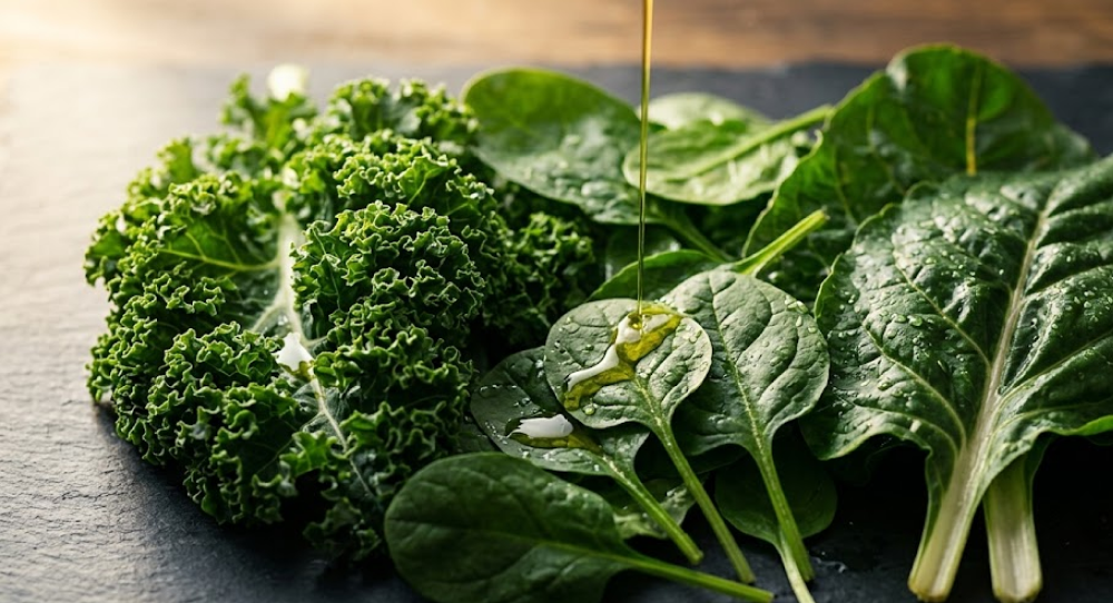
Optimizing daily consumption of dietary Vitamin K stands as one of the simplest and most impactful strategies for supporting baseline vascular security. The richest natural concentrations of Vitamin K1 reside inside dark green leafy vegetables, where the nutrient is stored directly within the photosynthetic machinery of plant cells. Consuming foods such as kale, spinach, Swiss chard, and collard greens provides abundant quantities of natural phylloquinone. However, because these nutrient molecules are tightly bound inside plant cell walls, raw consumption often yields relatively low absorption rates compared to gently cooked or properly paired culinary preparations.
Because phylloquinone functions as a classic fat-soluble nutrient, its uptake in the digestive tract relies heavily on dietary fats and healthy bile production. Combining green vegetables with wholesome lipids like extra virgin olive oil, avocado, or grass-fed butter stimulates digestive micelle formation. These microscopic lipid spheres transport Vitamin K efficiently across the intestinal lining and into the lymphatic system. Biohackers aiming for maximum bioavailability optimization should realize that eating dry raw salads results in significantly less nutrient absorption than consuming cooked greens paired with healthy fats. Mastering these practical meal composition habits ensures consistent systemic delivery without relying exclusively on synthetic supplementation.
- Dark green vegetables: Provides exceptionally dense natural sources of Vitamin K1 through spinach, kale, and collard greens.
- Dietary fat integration: Combines green vegetables with healthy fats to dramatically boost digestive micelle creation and uptake.
- Gentle culinary heating: Breaks down tough plant cell walls through light steaming or sautéing to release bound phylloquinone.

Vitamin K1 vs. Vitamin K2: Distinct Roles in Body Dynamics
While both molecules belong to the same overarching nutrient family, comparing Vitamin K1 vs K2 reveals vastly different physiological target destinations and biological duties. Vitamin K1 features a single saturated side chain, whereas Vitamin K2—known as menaquinone—contains a series of unsaturated isoprenoid units. These molecular structural variations dictate how the human body absorbs, distributes, and utilizes each form over time. Vitamin K1 is rapidly cleared from circulation by liver tissue to support liver targeted clotting factor synthesis, while Vitamin K2 remains in the bloodstream longer to reach peripheral tissues like bones and arterial walls.
Understanding these menaquinone differences enables biohackers to tailor their nutrient protocols without confusing biological outcomes. While Vitamin K1 serves as the body’s primary emergency sealant by maintaining hemostasis process capacity, Vitamin K2 activates osteocalcin and matrix Gla protein to govern calcium placement throughout the skeleton. This secondary function plays an essential role in arterial calcification prevention, ensuring that circulating calcium deposits into bone matrix rather than soft arterial tissue. Both nutrients work synergistically to maintain long-term cardiovascular health, but Vitamin K1 remains the non-negotiable molecular foundation for functional blood clot assembly.
- Tissue target destination: Directs Vitamin K1 primarily to hepatic tissue while Vitamin K2 travels to bones and arteries.
- Circulatory half-life duration: Clears K1 within hours to support rapid coagulation, whereas long-chain K2 forms persist for days.
- Primary physiological focus: Governs blood clotting factor activation with K1 while regulating systemic calcium movement with K2.
Final Thoughts on “Vitamin K1 and Clotting: The Body’s Natural Sealant”
Mastering foundational biochemistry is the true mark of intelligent health optimization, and understanding the role of Vitamin K as the body’s primary molecular sealant highlights this principle perfectly. While advanced biohacking protocols often focus on mitochondrial longevity or cognitive enhancement, neglecting basic hemostasis process efficiency compromises the vascular system’s structural integrity. Ensuring consistent daily intake of Vitamin K1 through dark leafy greens paired with healthy fats provides your liver with the necessary raw materials to synthesize fully functional clotting factors. By prioritizing whole-food phylloquinone sources, you establish a resilient defense mechanism that rapidly seals micro-trauma, limits unnecessary tissue inflammation, and protects long-term cardiovascular performance.
Ultimately, optimal health relies on maintaining harmony between biochemical supply and physiological demand. Fat-soluble Vitamin K represents an irreplaceable pillar of vascular resilience, bridging the gap between everyday nutrition and advanced physical recovery. Whether your goal is accelerating recovery from intense training, optimizing blood clotting mechanics, or simply supporting natural tissue repair, maintaining predictable daily micronutrient levels remains essential. Respecting the fundamental power of Vitamin K allows biohackers to build a truly robust health baseline, ensuring the body’s natural sealing system remains perpetually primed, reliable, and capable of executing flawless biological repairs whenever vascular emergency strikes.
- Foundational health baseline: Prioritizes basic hemostasis process stability before attempting complex biohacking or performance enhancement protocols.
- Nutritional consistency focus: Delivers steady daily amounts of natural Vitamin K1 through lipid-paired leafy green vegetables for reliable liver storage.
- Long-term vascular resilience: Maintains seamless blood clotting capabilities while safeguarding structural blood vessel integrity against unexpected micro-injuries.